Introduction:
Traumatic brain injuries (TBIs) can devastate an individual’s physical, cognitive, and emotional well-being. Assessing the severity of a TBI is crucial for determining appropriate medical interventions, predicting outcomes, and providing timely and targeted rehabilitation. This article aims to provide a guide to assessing the severity of a traumatic brain injury.
Please note that we do not recommend self-assessment of a TBI. If a TBI has impacted you or someone near you, please seek qualified medical professionals who can properly assess the severity of a TBI. Failure to properly assess a TBI can have severe negative consequences.
Glasgow Coma Scale (GCS)
The Glasgow Coma Scale is a widely used tool to assess the level of consciousness and neurological functioning after a TBI. The GCS score ranges from 3 to 15, with lower scores indicating more severe injuries. GCS scores can help classify TBIs into mild, moderate, or severe categories.
The total GCS score is the sum of the scores for each category.
- Eye-opening: The eye-opening category is scored based on the patient’s spontaneous eye-opening, eye-opening to verbal stimuli, and eye-opening to pain.
- Verbal response: The verbal response category is scored based on the patient’s ability to speak and their level of comprehension.
- Motor response: The motor response category is scored based on the patient’s ability to move their limbs in response to verbal commands and pain.
A score of 15 indicates that the patient is fully awake and alert, while a score of 3 indicates that the patient is in a coma. The lower the score, the more severe the brain injury or medical condition.
The GCS is a quick and easy way to assess the level of consciousness in patients with brain injuries or other medical conditions that can affect consciousness. Healthcare professionals in hospitals, emergency departments, and other healthcare settings use the scale.
Imaging Techniques Used in Assessing the Severity of TBI:
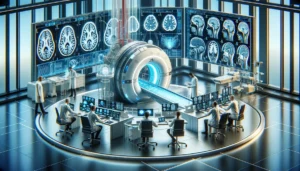
Various imaging techniques, such as computed tomography (CT) and magnetic resonance imaging (MRI), play a vital role in assessing the severity of a TBI. CT scans can quickly identify skull fractures, hemorrhages, and other acute abnormalities, while MRI scans provide detailed information about structural damage, such as contusions, hematomas, and diffuse axonal injury.
The two most common imaging techniques in assessing the severity of a TBI are computed tomography (CT) scans and magnetic resonance imaging (MRI) scans.
- CT scans use X-rays to create detailed images of the brain. CT scans are quick and easy to perform, making them ideal for the initial assessment of TBI patients. CT scans can identify skull fractures, blood clots, and other injuries requiring immediate medical attention.
- MRI scans use a strong magnetic field and radio waves to create detailed brain images. MRI scans are more sensitive than CT scans and can be used to identify more subtle injuries, such as axonal injury and diffuse brain injury. MRI scans are also better at showing the brain’s blood vessels, which can be important for assessing the risk of stroke.
In addition to CT and MRI scans, other imaging techniques that may be used to assess the severity of a TBI include:
- Cerebral angiography is a type of X-ray that uses a contrast dye to show the brain’s blood vessels. This technique can be used to identify blood clots and other vascular injuries.
- Single-photon emission computed tomography (SPECT) is a nuclear medicine imaging technique that uses a radioactive tracer to show blood flow in the brain. SPECT scans can assess the severity of brain injury and track the patient’s progress over time.
- Diffusion tensor imaging (DTI) is a type of MRI scan that can be used to measure the movement of water molecules in the brain. DTI scans can be used to assess the integrity of white matter tracts, which are important for communication between different parts of the brain.
The type of imaging used in assessing the severity of a TBI will depend on the patient’s clinical presentation and the information the healthcare team needs to make treatment decisions. In general, CT scans are used as the first-line imaging modality for TBI patients, while MRI scans provide more detailed information about the extent of the injury.
The results of imaging studies can also be used to classify the severity of a TBI. For example, a CT scan that shows a large blood clot in the brain would indicate a more severe injury than a CT scan that shows only minor brain swelling.
Clinical Assessment of TBI:
Clinical assessment involves thoroughly evaluating physical, cognitive, and behavioral symptoms associated with a TBI. Healthcare professionals assess factors such as headache intensity, nausea, dizziness, memory loss, confusion, changes in speech or vision, motor deficits, and emotional disturbances. The severity of these symptoms can help determine the extent of the injury.
Other clinical assessments that may be used to assess TBI include:
- The AVPU scale is a simple tool used to assess the patient’s level of consciousness. The AVPU scale has four categories:
- A = Alert
- V = Verbal
- P = Pain
- U = Unresponsive
- The Rancho Los Amigos Scale is a more comprehensive assessment tool used to assess the patient’s cognitive function and level of consciousness. The Rancho Los Amigos Scale has ten levels, with level 1 being the most severely injured and level 10 being the least injured.
- The National Institutes of Health Stroke Scale (NIHSS) is a neurological assessment tool used to assess the patient’s level of consciousness, motor function, and other neurological signs. The NIHSS has 15 items, with a higher score indicating a more severe injury.
Clinical assessments are an important part of the initial assessment of TBI patients. The information gathered from clinical assessments can be used to classify the severity of the injury, make treatment decisions, and monitor the patient’s progress over time.
Intracranial Pressure Monitoring:
In severe cases, monitoring intracranial pressure (ICP) becomes essential. Increased ICP can indicate the presence of brain swelling or bleeding, potentially leading to secondary brain injury. Intracranial pressure monitoring involves inserting a small catheter into the brain to measure pressure levels and guide treatment decisions.
Here’s how ICP monitoring is used in assessing the severity of a TBI:
Measurement of ICP:
ICP monitoring involves the placement of a monitoring device, such as an intraventricular catheter, intraparenchymal sensor, or subdural bolt, into the skull to directly measure the pressure inside the brain. The monitoring device is connected to a transducer, which converts the pressure into electrical signals that can be measured and displayed on a monitor.
Early Detection of Increased ICP:
TBI can lead to increased intracranial pressure, which can further damage the brain and impede proper blood flow. ICP monitoring allows for the early detection of elevated ICP levels, which may indicate worsening brain swelling, hemorrhage, or other complications. Sustained or rapidly rising ICP can suggest more severe brain injury.
Assessment of Treatment Efficacy:
ICP monitoring helps healthcare providers assess the effectiveness of interventions to reduce intracranial pressure. Medications, cerebrospinal fluid drainage, osmotic therapy, or surgical procedures can be monitored to determine their impact on ICP levels. If the ICP remains high despite interventions, it may indicate the need for more aggressive measures or neurosurgical interventions.
Detection of Critical ICP Thresholds:
Certain ICP thresholds are considered critical and may require immediate action to prevent further brain injury. For example, sustained ICP above 20-25 mmHg is typically considered dangerous, and measures to lower the pressure may be necessary. ICP monitoring helps identify these critical thresholds and guides treatment decisions.
Prognostic Value:
Persistent elevation of ICP or failure to control ICP within acceptable limits can be indicative of a more severe TBI and may be associated with a poorer prognosis. Monitoring ICP trends over time can assist in predicting the potential outcomes and long-term effects of the TBI.
It’s important to note that ICP monitoring is an invasive procedure typically reserved for patients with moderate to severe TBI or those at high risk for intracranial hypertension. The medical team decides to use ICP monitoring on a case-by-case basis, considering the patient’s clinical condition, imaging findings, and other factors.
Neuropsychological Assessment of TBI:
Neuropsychological testing helps evaluate cognitive function, memory, attention, language skills, and emotional well-being following a TBI. These assessments provide objective measurements and can track changes in cognitive abilities over time. Neuropsychological tests contribute valuable information for determining the severity of the injury and developing targeted rehabilitation plans.
Here’s how neuropsychological assessment is used to assess TBI severity:
Cognitive Functioning:
Neuropsychological tests assess a range of cognitive domains, including attention, memory, executive function, language, visuospatial skills, and processing speed. By evaluating these areas, the severity and specific patterns of cognitive impairment can be identified. For example, deficits in attention and memory may indicate a moderate to severe TBI, while milder deficits may suggest a mild TBI.
Comparison to Normative Data:
Neuropsychological assessment involves comparing an individual’s test performance to established normative data. These norms are based on the performance of healthy individuals of similar age, education, and background. By comparing the TBI patient’s performance to these norms, clinicians can determine how much the injury has affected their cognitive abilities.
Deficit Profile:
Neuropsychological testing helps identify specific cognitive deficits and patterns of impairment associated with TBI. For example, difficulties with attention, working memory, and processing speed may be indicative of frontal lobe damage, while problems with memory encoding and retrieval may suggest temporal lobe involvement. The pattern of deficits can provide insights into the severity and localization of the brain injury.
Functional Implications:
Neuropsychological assessment goes beyond identifying cognitive deficits; it also evaluates the functional implications of these deficits in everyday life. By examining the impact on activities of daily living (ADLs), social interactions, employment, and overall quality of life, clinicians can gain a better understanding of the functional limitations resulting from the TBI and the associated severity.
Longitudinal Assessment:
Neuropsychological testing can be performed at different stages of recovery to monitor the progression of cognitive functioning over time. This longitudinal assessment allows clinicians to track changes in cognitive abilities and determine the recovery trajectory or identify any persistent deficits.
Treatment Planning and Rehabilitation:
Neuropsychological assessment results play a crucial role in developing individualized treatment plans and rehabilitation strategies. By identifying specific cognitive strengths and weaknesses, clinicians can target interventions to address the areas of impairment and maximize recovery potential.
Neuropsychological assessment is typically conducted by trained neuropsychologists or clinical psychologists with expertise in brain injury assessment. The assessment battery may include standardized tests, questionnaires, interviews, and observations. The specific tests used may vary depending on the individual’s age, education level, cultural background, and suspected areas of impairment.
By integrating the findings from the neuropsychological assessment with other clinical evaluations, such as neurological examinations and neuroimaging, healthcare providers can understand the severity and functional impact of the TBI. This information aids in treatment planning, counseling, and providing appropriate support to individuals recovering from a TBI.
Biomarkers and Advanced Techniques:
Researchers are exploring using biomarkers in assessing the severity of a TBI. Biomarkers are measurable indicators found in blood, cerebrospinal fluid, or other bodily fluids that reflect the extent of brain damage. Additionally, advanced techniques like diffusion tensor imaging (DTI) and functional MRI (fMRI) offer insights into the structural and functional changes in the brain after a TBI.
While biomarker research in TBI is still evolving, several biomarkers have shown promise in assessing the severity of a TBI. Here’s an overview of their use:
- S100B: S100B is a calcium-binding protein primarily expressed in astrocytes. Elevated levels of S100B in the blood shortly after TBI have been associated with more severe injuries and worse clinical outcomes. It has been used as a biomarker to identify patients requiring further evaluation or neuroimaging.
- Neuron-Specific Enolase (NSE): NSE is an enzyme found primarily in neurons. Elevated levels of NSE in blood or CSF have been associated with neuronal damage in TBI. Higher NSE levels have been correlated with more severe brain injury and poor outcomes.
- Glial Fibrillary Acidic Protein (GFAP): GFAP is an intermediate filament protein in astrocytes. Increased levels of GFAP in blood or CSF have been observed after TBI. Elevated GFAP levels have shown associations with TBI severity and can help predict the presence of intracranial lesions on neuroimaging.
- Tau Protein: Tau is a microtubule-associated protein found in neurons. Elevated levels of tau in blood or CSF have been observed in TBI cases, particularly in more severe injuries. Tau has shown associations with white matter injury and may help predict long-term cognitive and functional outcomes.
- Neurofilament Light Chain (NFL): NFL is a protein component of the neuronal cytoskeleton. Increased levels of NFL in blood or CSF have been associated with axonal injury and neuronal damage in TBI. Higher NFL levels have shown correlations with TBI severity, acute injury characteristics, and long-term outcomes.
- Cerebrospinal Fluid Biomarkers: Biomarkers such as amyloid-beta (Aβ) peptides and tau protein in CSF have been investigated in the context of TBI. They have shown associations with neurodegenerative processes, including chronic traumatic encephalopathy (CTE), which can develop following repeated head injuries.
It’s important to note that while biomarkers have shown promise in TBI assessment, they are not yet routinely used in clinical practice. Further research is needed to establish standardized protocols, define cutoff values, and validate their clinical utility. Biomarker measurements are often used in conjunction with clinical assessments, neuroimaging, and other diagnostic tools to enhance the understanding of TBI severity and guide treatment decisions.
Biomarkers can also monitor the response to treatment, predict long-term outcomes, and contribute to research efforts to improve our understanding of TBI and develop targeted therapies.
Post-Concussion Symptom Scale:
The Post-Concussion Symptom Scale (PCSS) is a self-reporting tool used to assess the severity and duration of post-concussion symptoms. Patients rate the intensity of symptoms such as headache, dizziness, fatigue, sleep disturbances, and emotional changes. The PCSS aids in evaluating the progression of symptoms and determining when an individual can safely return to normal activities.
The PCSS captures a broad range of physical, cognitive, emotional, and sleep-related symptoms commonly associated with concussions. Here’s an overview of the PCSS:
- Symptom Assessment: The PCSS includes a list of 22 symptoms commonly reported after a concussion. These symptoms include headaches, dizziness, fatigue, sleep disturbances, memory difficulties, irritability, anxiety, and sensitivity to light or noise.
- Symptom Severity: For each symptom, the patient rates its severity on a scale ranging from 0 to 6, with 0 indicating no symptom and 6 indicating the most severe symptom. The severity ratings provide a quantitative measure of symptom intensity.
- Symptom Frequency: The PCSS also captures the frequency of each symptom experienced by the patient over the past 24 hours. The patient rates the frequency on a scale ranging from 0 to 6, with 0 indicating no symptom and 6 indicating the symptom being experienced constantly.
- Symptom Total Score: The PCSS calculates a total score by summing up the severity ratings across all the symptoms. This total score provides an overall measure of symptom burden and can be used to track changes in symptom severity over time.
- Symptom Monitoring: The PCSS is administered at various time points following a concussion, such as immediately after the injury, within the first week, and at subsequent follow-up appointments. By comparing scores across different time points, healthcare providers can assess the trajectory of symptom recovery and identify any persistent or worsening symptoms.
- Individualized Tracking: The PCSS allows patients to track their symptoms and communicate their experiences with healthcare providers. Patients can note changes in symptom severity or frequency, providing valuable information for managing their recovery.
The PCSS is a widely used tool for both research and clinical purposes. It helps healthcare providers objectively assess and monitor post-concussion symptoms, guide treatment decisions, and facilitate communication between patients and healthcare teams. However, it’s important to note that the PCSS is just one component of a comprehensive evaluation, and other assessments, such as neurological examinations and neuropsychological testing, may be necessary to evaluate the full extent of the injury and its impact on cognitive functioning and overall well-being.
Conclusion:
Assessing the severity of a TBI requires a multidimensional approach. Utilizing tools like the Glasgow Coma Scale, imaging techniques, clinical assessments, intracranial pressure monitoring, neuropsychological evaluations, biomarkers, and self-reporting scales provides a comprehensive understanding of the injury’s extent and guides treatment decisions. A timely and accurate assessment is crucial for optimizing patient care, predicting outcomes, and developing tailored rehabilitation plans to improve long-term outcomes for individuals with traumatic brain injuries.
The severity of a TBI is an important factor in determining the patient’s prognosis. Patients with mild TBIs typically fully recover, while patients with severe TBIs may have long-term disabilities. If you or someone close has experienced a TBI, then please seek help from qualified medical professionals.


