Electroencephalography, commonly known as EEG, is a pivotal method in neuroscience and neurology for recording the brain’s electrical activity. This technique involves using electrodes placed on the scalp to detect and record the brain’s electrical impulses. The origins of EEG trace back to the early 20th century, with significant contributions from Hans Berger, a German psychiatrist who first demonstrated the human brain’s electrical activity. Since then, EEG has become an indispensable tool for research and clinical practices, providing insights into brain functions and diagnosing neurological disorders.
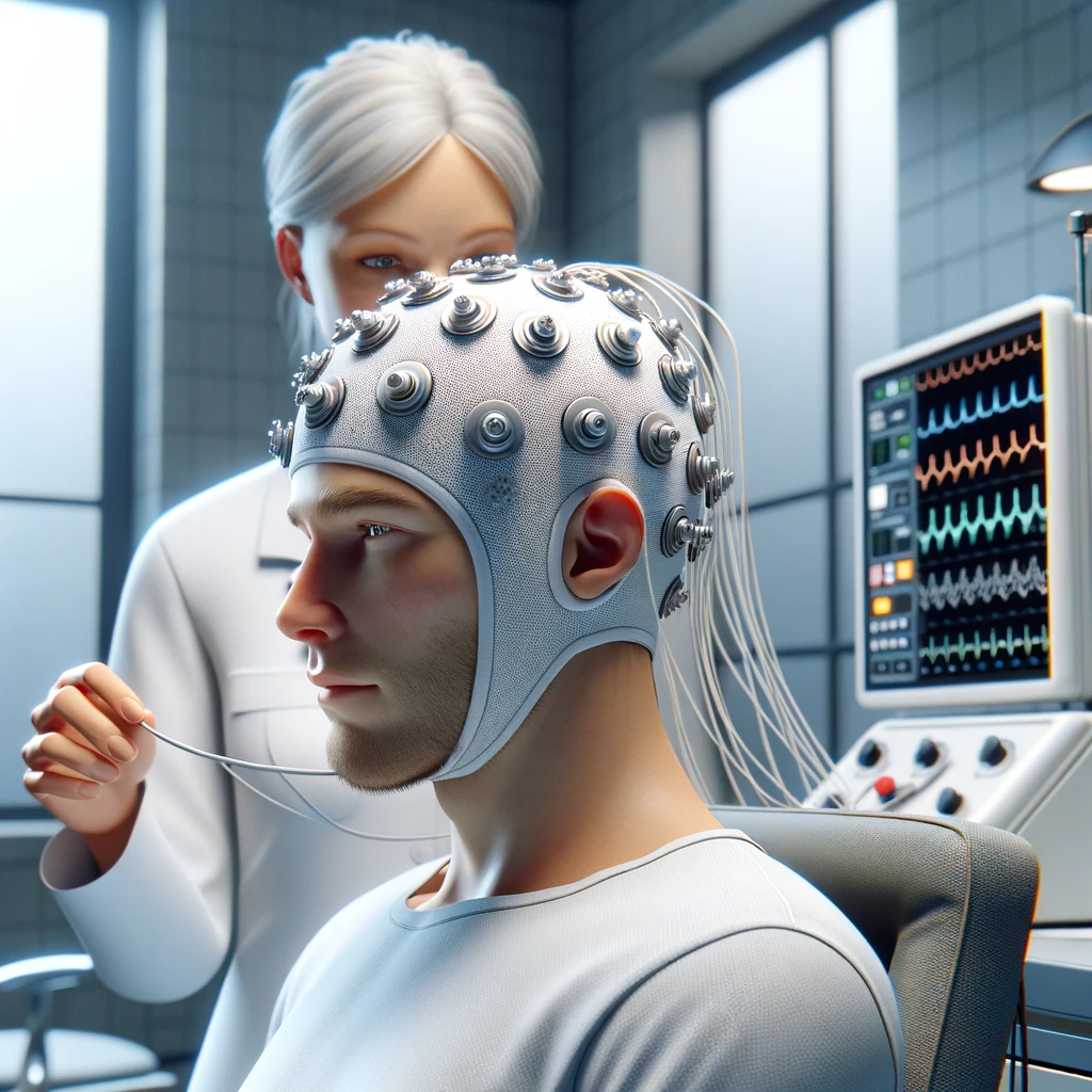
The process of conducting an EEG involves the placement of multiple electrodes at specific locations on the scalp. These electrodes capture the electrical signals generated by the neurons in the brain. A crucial aspect of EEG is using a standardized placement system, such as the International 10-20 system, which ensures consistency and comparability of recordings across different sessions and subjects. The electrodes connect to an amplifier and a recording device, which processes and records the signals. The recorded data, often represented as waveforms, reflect the brain’s electrical activity over time, allowing for detailed analysis.
EEG’s capability to provide real-time monitoring of brain activity makes it exceptionally valuable for clinical and research purposes. In addition, Doctors utilize EEG to diagnose and monitor neurological disorders such as epilepsy, sleep disorders, and brain injuries. EEG’s ability to detect abnormalities in brain wave patterns is crucial in identifying epileptic seizures, distinguishing different types of seizures, and guiding treatment plans. Moreover, EEG is a critical tool in monitoring the depth of anesthesia during surgical procedures, ensuring patient safety.
Continuing Research for using EEG for Assessment of TBI
In the research domain, EEG offers a window into understanding the brain’s functioning and neural mechanisms underlying various cognitive processes, such as attention, memory, and language. Researchers value EEG for its high temporal resolution, which allows them to observe how the brain’s activity changes over milliseconds, offering insights into cognitive processes’ dynamics. Furthermore, EEG studies contribute to the development of brain-computer interfaces (BCIs), enabling individuals to control devices or communicate through the modulation of their brain activity.
Advantages and Disadvantages
One of EEG’s most significant advantages is its noninvasive nature. Unlike other imaging techniques that may require exposure to radiation or the injection of contrast agents, EEG poses minimal risk to the participants, making it suitable for repeated use in adults and children. In addition, this aspect is particularly beneficial in longitudinal studies, where researchers must monitor brain activity over time.
Despite its advantages, EEG has limitations, primarily in its spatial resolution. The skull and scalp diffuse the electrical signals, making it challenging to localize the source of brain activity precisely. Advanced techniques, such as source localization methods, aim to overcome this limitation by combining EEG data with information from other imaging modalities, such as MRI, to enhance the accuracy of identifying the brain regions involved in generating the recorded signals.
EEG for TBI Assessment
Electroencephalography (EEG) plays a significant role in the assessment and management of traumatic brain injury (TBI), offering valuable insights into the functional consequences of brain injuries. The severity of a TBI, ranging from mild to severe, can have profound implications on a patient’s treatment, recovery, and prognosis. EEG for Assessment of a TBI contributes by providing real-time information on brain activity, which can help identify disturbances caused by the injury. Here are several ways EEG aids in determining the severity of a traumatic brain injury:
1. Detection of Abnormal Brain Activity
EEG is highly sensitive to abnormalities in brain electrical activity. After a TBI, the brain may exhibit unusual patterns such as slow waves (indicative of brain dysfunction or damage), sharp waves, or seizures. The presence and distribution of these abnormalities can help clinicians assess the extent and impact of the injury.
2. Assessment of Brain Function
The overall brain function, as reflected in the EEG, can provide critical information on the severity of the injury. For example, a diffusely slow EEG pattern might indicate a moderate to severe brain injury, reflecting widespread brain dysfunction. In contrast, a routine EEG might suggest a milder form of TBI, although it’s essential to consider that a normal EEG does not rule out the presence of a TBI.
3. Monitoring for Seizure Activity
TBIs can lead to the development of seizures or epilepsy, which may not be immediately apparent. Continuous EEG monitoring is crucial for detecting non-convulsive seizures, which are common in individuals with moderate to severe TBIs. Identifying and treating these seizures promptly is vital for preventing further brain damage and improving outcomes.
4. Evaluating Consciousness Levels
EEG plays a role in assessing the level of consciousness in patients with severe TBIs, particularly those in comas or with altered mental states. Consequently, the EEG patterns can provide insight into the depth of coma and the potential for recovery. For example, specific EEG patterns, like reactivity to stimuli, may indicate a better prognosis.
5. Prognostic Indications
Specific EEG patterns can have prognostic significance. For instance, when using EEG for TBI assessment, the persistence of abnormal slow-wave activity can suggest a more extended recovery period or a higher likelihood of lasting cognitive impairment. Conversely, the normalization of EEG patterns over time can indicate recovery of brain function.
6. Guiding Rehabilitation and Treatment
EEG can help tailor rehabilitation strategies by providing insights into the brain’s functional status. In cases of severe TBI, EEG may assist in decisions regarding the intensity of rehabilitation efforts or the need for interventions to address specific neurological deficits.
It’s important to note that while EEG is a powerful tool for assessing brain activity, determining the severity of TBI typically involves a comprehensive evaluation, including clinical examination, neuroimaging techniques like MRI or CT scans, and neuropsychological assessments. Furthermore, EEG adds a crucial dimension by offering a dynamic view of brain function, complementing other diagnostic modalities to provide a more rounded understanding of the injury’s impact.
In conclusion, electroencephalography is a cornerstone in exploring the brain’s electrical activity, offering profound insights into the human brain’s functioning. Its applications span clinical diagnostics to cutting-edge research, reflecting its versatility and importance in neuroscience. Despite its limitations, ongoing advancements in EEG technology and analysis methods continue expanding its capabilities, promising even more extraordinary contributions to our understanding of the brain and advancing neurology and cognitive sciences.