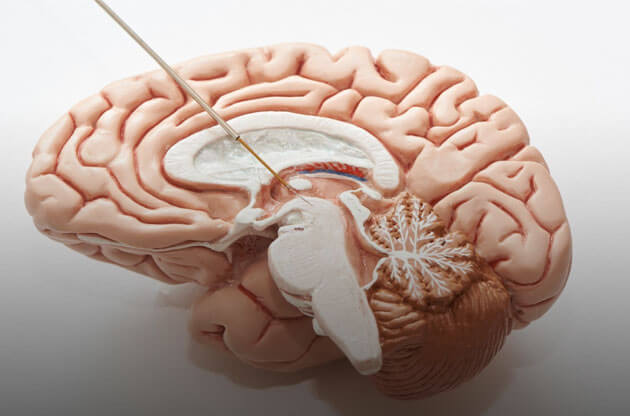
Even in modern times, traumatic brain injury (TBI) is still the foremost cause of morbidity and mortality. With various experimental and clinical studies, we have expanded our knowledge of traumatic brain injury, its pathophysiology, and treatment strategies.
Pathophysiology of traumatic brain injury
The initial stages of traumatic brain injury are characterized by tissue damage. Some primary pathophysiological events may trigger secondary brain injury over time. Traumatic brain injury leads to impaired regulation of cerebrospinal fluid and metabolism. This further leads to lactic acid accumulation, increased permeability, and the formation of oedema.
Neurochemical changes: Energy-dependent sodium-potassium ATPase pumps regulate ionic changes across the membrane. Traumatic brain injury disrupts the cell membrane that induces the redistribution of ions and neurotransmitters. In the further events of traumatic brain injury pathophysiology, this depolarization of the terminal membrane occurs along with the excessive release of glutamate and aspartate (excitatory neurotransmitters), voltage-dependent calcium and sodium channels, etc. The calcium and sodium influx leads to carbolic intracellular processes, increasing the intracellular concentration of free radicals and free fatty acids. Besides, there occurs activation of caspases, endonucleases, and translocate, which further initiates progressive changes in the structure of biological membranes and the nucleosomal DNA. Ultimately, these events lead to programmed or necrotic cell death.
Cerebral blood flow: Following a traumatic brain injury, hemodynamic and metabolic changes are specifically observed. Pathological hemodynamic changes emphasize brain dysfunction and cerebral ischemia. Hypotension, hypoxia, and collapse of cerebral blood flow (CBF) are observed following Traumatic brain injury. The hemodynamic status shows lethal hypotension levels, traumatized brain tissue, and deterioration of cerebral blood flow. Some patients develop cerebral hyperperfusion or hypoperfusion in the early stages of injury. Also, the risk of hyperaemia post-traumatic ischemia is high. This pathology is damaging as it may cause vasoparalysis.
Cerebrovascular autoregulation and CO2-reactivity: These two are regulatory patterns and manage cerebral perfusion pressure (CPP) and intracerebral pressure (ICP). Any impairment in these mechanisms shows a high risk for secondary brain damage. Following traumatic brain injury, cerebrovascular autoregulation is impaired. Traumatic brain injury pathophysiology of the temporal profile shows inconsistency as the severity of the injury. This kind of defective cerebrovascular autoregulation may occur immediately after the injury or may develop later. The cerebrovascular CO2-reactivity is, however, a robust phenomenon. In cases of severe brain injury, CO2-reactivity is impaired in the initial stages after trauma.
Cerebral vasospasm: Vasospasm is common, and it indicates severe brain damage. Traumatic brain injury pathophysiology shows vasospasm occurs by many mechanisms, including chronic depolarization of vascular smooth muscle, the release of endothelin, reduced availability of nitric oxide, and depletion of cyclic GMP of vascular smooth muscle, free radical formation, etc.
Cerebral oxygenation: There is an imbalance between delivery and consumption of cerebral oxygen following a traumatic brain injury, leading to brain tissue hypoxia.
Cerebral metabolic dysfunction: Consumption of cerebral oxygen and glucose reflects cerebral metabolism. After traumatic brain injury, cerebral metabolism and cerebral energy decrease. The degree of metabolic dysfunction relates to the severity of the trauma. Post-traumatic cerebral metabolism reduction relates to the initial trauma leading to mitochondrial dysfunction and intramitochondrial calcium overload. Sometimes, an alternative pathophysiological event like hypermetabolism of glucose can occur.
Excitotoxicity and oxidative stress: Following traumatic brain injury, there is an excessive release of excitatory amino acid neurotransmitters like glutamate, which act as neurotoxins that can kill nerve cells. Traumatic brain injury pathophysiology features persistent depolarization of neuronal cells and astrocytes. This leads to the over-stimulation of ionotropic and metabotropic glutamate receptors with consecutive sodium, potassium, and calcium fluxes. To restore normal ionic gradients, the brain demands production of energy for restoring ionic homeostasis. But because of inadequate cerebral blood flow, mitochondrial dysfunction occurs, and ultimately, it results in excitotoxic neuronal cell damage and death. Similarly, oxidative stress because of excitotoxicity results in peroxidation of vascular and cellular structures, cleavage of DNA, protein oxidation, and inhibition of the mitochondrial electron transport chain. They contribute to inflammatory processes and immediate cell death.
Oedema: Any structural, water, and osmotic imbalance induced by traumatic brain injury is known as ‘brain oedema’. Vasogenic brain oedema and cytotoxic is commonly seen in patients after the injury.
Inflammation: In traumatic brain injury pathophysiology, the release of cellular mediators such as prostaglandins, proinflammatory cytokines, and free radicals is very common.
Cell death: In cell death, many groups of biochemical pathways and proteins are involved. In necrosis cell membranes get damaged, and there is an uncontrolled release of cell death. However, apoptosis (a naturally programmed cell death) is triggered by DNA damage, cell surface receptor engagement, and withdrawal of growth factors.
Thus, traumatic brain injury pathophysiology is a complex process that starts many events of pathological cellular pathways. With a better understanding of neurochemical and metabolic responses to Traumatic brain injury pathophysiology, tailored treatment can be offered to patients.