The management of traumatic brain injury (TBI) is complex and presents many challenges. In most cases of TBI, patients also have injuries to other organ systems. This further complicates its management. Mass lesions from brain injury need surgical evacuation as it is a life-saving measure. Surgical management of TBI also improves outcomes and is commonly used to remove intracranial hemorrhage (ICH).
The surgical management of traumatic brain injury depends on the type of injury and the neurologic examination. Traumatic parenchymal lesions, acute epidural hematoma (aEDH), acute subdural hematoma (aSDH), posterior fossa mass lesions, large vessel injuries, depressed cranial fractures, etc., require surgical consideration.
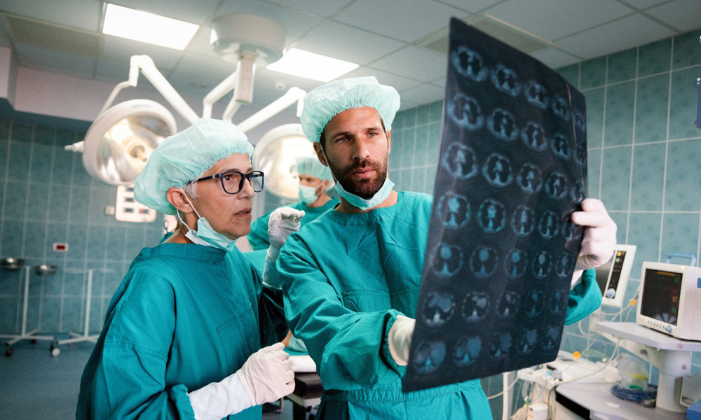
Surgical management
ICP monitoring is essential for both medical and surgical decision-making. It helps to understand cerebral perfusion and functions as intracranial compliance. Hence, ICP monitoring is performed in all salvageable patients.
Acute epidural hematoma (aEDH): Loss of consciousness, lucid interval and obtundation, and a lens-shaped hemorrhage on computed tomography (CT) is a presentation of aEDH. It occurs in the temporal and temporoparietal regions. The source of hemorrhage is a linear skull fracture with damage to the middle meningeal artery. Surgical management of this type of hematoma is based on imaging and patient status. aEDH larger than 30cc needs surgical intervention regardless of the patient’s Glasgow Coma Scale (GCS) score. Expeditious hematoma evacuation is important for comatose patients (GCS < 9) with anisocoria. With the advancement in imaging quality and technology, surgeons can localize the clot before incision, allowing for a smaller linear incision.
Acute subdural hematoma (aSDH): Indications for surgery for aSDH is a thickness greater than 10 mm and midline shift over 5 mm on the CT scan. All the traumatic brain injury patients with such findings are operated regardless of their GCS. The preferred method of surgical management of aSDH is a craniotomy with or without bone flap removal or duraplasty. The method depends on intraoperative findings.
Acute SDH patients with GCS < 9 should have intracranial pressure monitoring. For patients in a coma, surgical evacuation is indicated with ICP > 20mmHg, less than a 10mm thick lesion or less than 5mm of midline shift, a decrease of GCS score by two or more points, or asymmetric or fixed and dilated pupils.
Traumatic parenchymal lesions: Surgical management is needed if there is a progressive neurological deterioration with the intracerebral lesion. Patients with GCS 6-8 with contusions greater than 20 cm3 and minimum of 5mm of midline shift or cisternal compression on CT scan need surgical management. The surgical intervention method depends on the nature and location of the injury. A craniotomy is considered for traumatic parenchymal lesion indications. For patients suffering from medically refractory intracranial hypertension, surgeons may consider a bifrontal decompressive craniectomy (DC). A temporal lobectomy, hemispheric DC, and subtemporal decompression are other options for decompression.
Depressed cranial fractures: Skull fractures may warrant surgical management in cases such as open (compound) cranial fractures. In these types of fractures, the fractures are depressed greater than the thickness of the cranium. Hence, it needs surgery to prevent infection and any further damage to the brain parenchyma. Sometimes, open (compound) depressed cranial fractures are treated nonoperatively if there is no radiographic or clinical evidence of dural penetration, intracranial hematoma, frontal sinus involvement, depression greater than 1 cm, wound infection, or contamination. In such cases, early surgical intervention like elevation and debridement is advised to decrease the chance of infection. In the absence of wound infection, primary bone fragment replacement is a good surgical option. The management for open (compound) depressed fractures must include anti-epileptic drugs and antibiotics.
Penetrating trauma: It is a unique area of traumatic brain injury that occurs because of various objects, weapons such as firearms. In such cases, antibiotics and antiepileptic drugs are given as soon as possible. The primary focus of surgical management of penetrating trauma is to control ICPs, secondary injury, and the removal of the penetrating foreign object.
Several surgical management strategies can help a patient with traumatic brain injury in coping with the complications.