Our brain is protected by the skull’s bony protection, which prevents any serious harm to the brain. Head injuries are caused by a blow to the head and are usually associated with motor vehicle accidents, falls, physical assaults, or any sport-related accidents. In case of a severe head injury, the damage may affect the brain, and in the worst case, it may even affect the spine. There are different types of severe head injuries based on the effect it has on the head or brain including,
- Hematoma: It includes collecting of blood outside the blood vessels inside the brain. The accumulation can increase pressure inside the skull.
- Hemorrhage: Hemorrhage because of severe head injury leads to uncontrolled bleeding in the area around the brain. If not controlled, it may lead to brain damage.
- Concussion: In this type of head injury, there is a severe impact on the head, which leads to brain injury.
- Edema: It causes swelling of the surrounding tissues in the brain. This increases pressure in the brain and causes the brain to press against the protective skull.
- Skull fracture: A broken skull cannot absorb the impact of a blow, making it more likely to cause damage to the brain.
- Diffuse axonal injury: Diffuse axonal injury causes no bleeding, but it results in damage to the brain cells and brain function loss.
Methods for screening and detection of severe head injury
Several methods are used to screen or detect the severity of the head injury. Based on these tests, doctors can determine how much damage the injury has caused to the brain. The different methods used for screening and detecting severe head injury include the following.
- Military Acute Concussion Evaluation (MACE): The MACE 2 is the screening test done immediately post-injury to determine cognitive deficits because of mild traumatic brain injury. It includes four cognitive domains tested for orientation, immediate memory, concentration, and delayed recall.
- Glasgow Coma Scale (GCS): This is one of the initial screening or detection methods used to determine the level of brain damage. The GCS is a 15-point test that helps to assess the patient’s mental status. A high GCS score indicates a mild injury. The GCS scale score can range from 3, which means completely unresponsive, to the scale of 15, which means the patient is responsive. In this screening method, the patient is examined for trauma signs, including bruising and swelling. The medical experts carry out a neurological examination to evaluate the nerve function by assessing muscle control, strength, eye movement, and sensation.
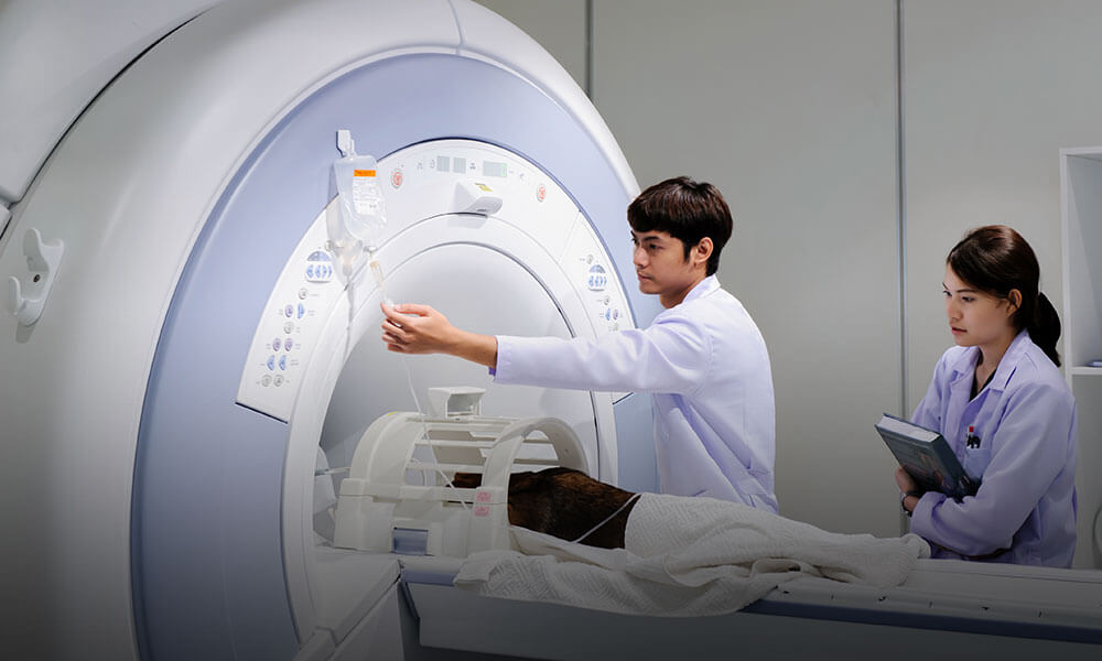
- Magnetic Resonance Imaging (MRI) scan: The MRI scans give information regarding the organ and the changes associated with ballistic trauma to the body. It provides the idea about the changes in the brain after the injury. The MRI scan is one of the non-invasive measurements carried out to check the severe head injury level. It helps to understand the brain contusion, which is the rupture of brain tissue or blood vessel, brain edema, vasospasm which include narrowing of the small arteries of the brain that leads to a hematoma, and hemorrhage in which it can help to detect the lesions caused in the brain because of severe brain injury.
- Neuronal Architecture Imaging methods: In case of a severe head injury, there are chances that along with the brain, there may be damage to the brain stem and the spinal cord. This method of screening will help to detect the damage caused to the corpus callosum and brain stem. The diffusion tensor imaging (DTI) method helps to detect damage to axonal tracts using a measure of directional water diffusion.
- Computerized Tomography (CT) scan: A CT scan is the most common imaging technique used to assess severe head injury because it readily detects trauma-related fractures, swelling of brain tissue, extra-axial fluid collection, hemorrhage, intracranial injury, and radio-opaque foreign bodies.
- PET and SPECT imaging: Positron emission tomography (PET) and Single Photon Emission Computerized Tomography (SPECT) scans are sensitive measurement methods. They help to understand brain metabolism and neuroreceptor concentrations after the injury. The different pathophysiological parameters imaged by PET include oxygen utilization, regional glucose metabolism, regional blood flow, and vasospasm detection, permeability, neuroreceptor concentrations, inflammation, beta-amyloid deposits associated with dementia, and Tau protein associated with brain trauma and dementia. These parameters change because of severe head injury.
- Ultrasound for brain blood flow: Measurements of blood flow in the brain basal arteries and the carotids by transcranial Doppler are effective methods for detecting vasospasm. The severe head injury results in a change in the flow characteristics.
The screening and detection techniques for severe head injury can help to detect the severity and the type of damage to the brain, and based on the results, the patient can get appropriate treatment.