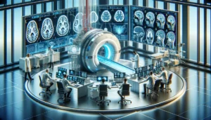
Traumatic brain injury (TBI) is a complex and potentially devastating medical problem. Even a mild injury may later develop into a serious clinical issue. Severe and moderate brain injuries are life-threatening and demand immediate medical attention. Your doctor will order high-sensitivity blood tests or TBI brain scans to rule out brain hemorrhage and contusion. Doctors suggest TBI brain scans depending on the nature, location, and severity of the injury.
TBI scans and tests
Neuroimaging scans and tests determine the presence and extent of traumatic brain injury. They help with surgical planning and specific interventions. TBI brain imaging is also critical in chronic therapy after the injury, identifying sequelae, determining prognosis, and rehabilitation guidance.
The scans and tests are categorized into two groups.
- The first group comprises tests that help to examine the structure and the function of the brain. It includes scans like the MRI and CT scan.
- Tests like SPECT scan, PET scan, EEG, evoked studies, etc., fall into the second group. They are widely used after a traumatic brain injury.
Some common TBI brain tests and scans include
- X-rays: An X-ray machine directs harmless X-rays on the head. A detector creates images of the radiation that passes through the head at different rates depending on the tissue density. An X-ray helps to visualize damage caused to the bones, like skull fractures.
- Computerized Tomography (CT): A CT scan is also known as computerized axial tomography (CAT). It uses a series of X-rays to create detailed images of internal areas of the head. Healthcare professionals use CT scans as the first test after the traumatic brain injury. It shows internal swelling, bleeding in the brain, and even a skull fracture. A CT scan creates multiple images of the head from different angles to create a detailed 3D internal image of the brain and the skull. The results of the scan are then used to identify the extent and exact location of the injury. This scan offers plenty of information for further tests and treatment. CT scans are also used for follow-ups during the recovery to monitor injury.
- Magnetic Resonance Imaging (MRI): A MRI scan machine uses magnetic fields and radio waves to create detailed computerized images of internal parts of the head from various angles. This diagnostic test does not expose the patient to X-rays. MRI scans are high-resolution pictures of brain structures. Also, an MRI scan is used to plan and to check the effects of the treatment.
- Positron emission tomography (PET): It requires advanced preparation and involves administering a radiotracer to the patient. This tracer emits low levels of radiation that the scanner detects and creates very detailed 3D pictures of activity in the brain. A combination scan is frequently used in TBI brain scans. PET scans are used in combination with CT or MRI scanners. This helps to take better and clearer images of brain tissue after a traumatic brain injury. Also, PET scans aids in diagnosing epilepsy or disorders of consciousness.
- Electroencephalography (EEG): Our brain cells communicate with each other and process information through electrical signals. An EEG detects the areas of brain activity and measures natural electrical activity in the brain. Electrodes are placed on the scalp surface to measure brain activity. This test can detect and monitor any abnormal brain activity after a traumatic brain injury. An EEG offers information on the effects of brain injury, like problems with sleep and memory.
- Single-Photon Emission Computed Tomography (SPECT): Like PET scans, a radiotracer is injected during the scan. The tracer provides 3D images of internal parts of the head. However, the radiotracer used in SPECT remains inside the body for a longer duration as compared with the radiotracer used in PET. TBI brain scan using SPECT technology helps to visualize injured brain regions and detect areas of decreased blood flow in the brain. Further, these scans offer information to plan surgeries and long-term treatments. As SPECT scans are specialized, they are not used frequently.
- Functional Magnetic Resonance Imaging (fMRI): It is a type of MRI scan but offers information about brain activity by measuring blood flow. It can identify the areas of the brain that are active under different conditions. TBI brain fMRI scans investigate disorders of consciousness and help in planning long-term treatments.
- Lumbar puncture: This is a diagnostic test in which cerebrospinal fluid is removed for examination, and the spinal column pressure is measured. It checks for the primary or metastatic brain, cerebral hemorrhage, or spinal cord neoplasm in cases of traumatic brain injury.
- Intracranial Pressure Monitor: As brain swelling is very dangerous, doctors insert an intracranial pressure monitor into the skull. It makes sure there is no elevated pressure that could worsen the traumatic brain injury.
- Magnetic Resonance Spectroscopy (MRS): It is another imaging method of detecting and measuring the brain’s activity at the cellular level. TBI brain MRS scans offer chemical information and are typically used with MRI.


